
What is OCT(Retina/Glaucoma)?
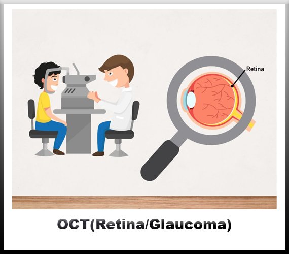
Optical Coherence Tomography (OCT) is a non-invasive imaging technique utilized in ophthalmology to visualize the layers of the retina, optic nerve head, and anterior segment of the eye. It provides high-resolution cross-sectional images, allowing clinicians to assess various eye conditions, including those related to the retina and glaucoma.
Retina refers to the light-sensitive layer of tissue lining the inner surface of the eye. It plays a crucial role in converting light into neural signals that are then transmitted to the brain, enabling vision. OCT imaging of the retina provides detailed information about its thickness, integrity, and any abnormalities present.
In retinal diseases such as age-related macular degeneration (AMD), diabetic retinopathy, and retinal detachment, OCT aids in early detection, monitoring
disease progression, and evaluating the effectiveness of treatments. For instance, in AMD, OCT can detect characteristic changes such as drusen deposits, retinal pigment epithelial detachment, and choroidal neovascularization.
Glaucoma is a group of eye conditions characterized by damage to the optic nerve, often associated with elevated intraocular pressure (IOP). This damage can lead to vision loss and, if left untreated, blindness. OCT imaging of the optic nerve head and retinal nerve fiber layer helps in diagnosing and managing glaucoma by assessing structural changes indicative of nerve damage, such as thinning of the retinal nerve fiber layer and excavation of the optic nerve head.
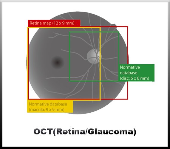
In OCT imaging for glaucoma, parameters such as the thickness of the retinal nerve fiber layer (RNFL) and the optic nerve head (ONH) are measured and compared to normative databases to detect early signs of glaucomatous damage. Additionally, OCT angiography, a recent advancement in OCT technology, allows for the visualization of retinal blood vessels and assessment of ocular perfusion, which is relevant in understanding the vascular component of glaucoma.
The integration of OCT into clinical practice has revolutionized the management of retinal and glaucomatous diseases by providing detailed anatomical information in a non-invasive manner. Its ability to detect subtle changes in retinal and optic nerve morphology enhances diagnostic accuracy, facilitates treatment decision-making, and enables monitoring of disease progression over time.
OCT technology continues to evolve, with advancements such as swept-source OCT and adaptive optics OCT further enhancing imaging capabilities and expanding clinical applications. These innovations hold promise for improving our understanding of ocular diseases and optimizing patient care in the field of ophthalmology.


Frequently Asked Questions
Choose Jain Eye Hospital for expert eye care. With cutting-edge technology and compassionate specialists, we ensure optimal vision health for you and your loved ones.
Over time, cataracts will harm your vision. Cataract surgery can bring back your vision. However, a possible complication of cataract surgery is an "after-cataract." An "after-cataract" happens when part of the natural lens that is purposely not taken out during cataract surgery becomes cloudy and blurs your eyesight.
How painful is cataract surgery? Most people feel little or no pain during or after cataract surgery. You'll receive a topical anesthetic (eye drops) to numb your eye during the surgery. Shortly after surgery, your eye may feel gritty or slightly tender, but over-the-counter pain medicine should improve this.
Many factors can induce inflammation of ocular surface, including DED, pathogens, and allergic reactions. Acute inflammation is usually beneficial, as it promotes the process of healing. But chronic inflammation may result in cell damage or even cell death.
Diagnosis and treatment of inflammatory eye diseases are important. They can cause permanent damage to the eyes and vision loss that cannot be reversed. If you notice any of the signs or symptoms of inflammatory eye disease, make an appointment to see your eye doctor right away for a complete eye exam.
We are the best
Welcome to Jain Eye Hospital, where your vision is our priority. With a legacy of excellence spanning over three decades, we are committed to providing world-class eye care services with compassion, expertise, and cutting-edge technology.
- expert in eye care.
- State-of-the-art facilities.
- Comprehensive services.
- Patient-centered approach.
Our Services


Diabetic Retinopathy Management
Such as photocoagulation or panretinal photocoagulation, halts blood and fluid leakage and shrinks abnormal vessels in the eye.
Read More
Ocular Inflammation
Ocular inflammation is inflammation of the eye, which can be caused by many factors, including pathogens, allergic reactions, and DED.
Read More
Visual Field Analysis
A visual field test, also known as a perimetry test, is a painless eye exam that measures the area of vision and how sensitive it is in different parts.It can detect blind spots in your vision.
Read More
We are available 24/7
We are Always Ready For A Challenge.
Satisfied Patients
Patient satisfaction is an attitude. Though it does not ensure that the patient will remain loyal to the doctor or the hospital, it is still a strong.
Awards
Highly skilled medical professionals offering specialized care and advanced treatment modalities.
How do you OCT(Retina/Glaucoma)?
Optical Coherence Tomography (OCT) has revolutionized the field of ophthalmology, particularly in the diagnosis and management of retinal and glaucoma diseases. This non-invasive imaging technique provides high-resolution cross-sectional images of ocular tissues, enabling clinicians to visualize the layers of the retina and optic nerve head with remarkable detail.
In OCT imaging of the retina, a patient is typically positioned comfortably in front of the OCT machine, and their eye is fixated on a target while the device captures scans using low-coherence interferometry. The OCT system emits infrared light, which penetrates ocular tissues and is reflected back from different layers of the retina. By analyzing the interference patterns between the reflected light and reference light, the OCT system generates detailed cross-sectional images of the retina.
These images allow clinicians to assess the thickness and integrity of retinal layers, identify abnormalities such as macular edema, epiretinal membranes, and macular holes, and monitor disease progression in conditions like diabetic retinopathy and age-related macular degeneration. Additionally, OCT angiography can provide information about retinal vasculature, aiding in the evaluation of conditions such as retinal vascular occlusions and neovascularization.
In OCT imaging of the optic nerve head for glaucoma evaluation, the focus is on assessing the structural integrity of the optic nerve fiber layer (ONFL) and measuring the thickness of the retinal nerve fiber layer (RNFL). Glaucoma is characterized by progressive damage to the optic nerve, often resulting in thinning of the RNFL. OCT allows for precise measurement of RNFL thickness, which can aid in early detection, monitoring disease progression, and assessing the effectiveness of treatment interventions.
During OCT imaging for glaucoma, the patient's pupil may be dilated to ensure optimal visualization of the optic nerve head and surrounding structures. The OCT scan captures cross-sectional images of the optic nerve head and peripapillary retina, providing detailed measurements of ONFL and RNFL thickness. These measurements are compared to normative databases to assess for abnormalities indicative of glaucomatous damage.
OCT imaging plays a crucial role in the diagnosis, management, and monitoring of both retinal and glaucoma diseases. Its non-invasive nature, high resolution, and ability to provide quantitative measurements make it an invaluable tool for ophthalmologists in clinical practice. As technology continues to advance, OCT is likely to become even more integral in the comprehensive evaluation of ocular pathology, contributing to improved patient outcomes and quality of care.