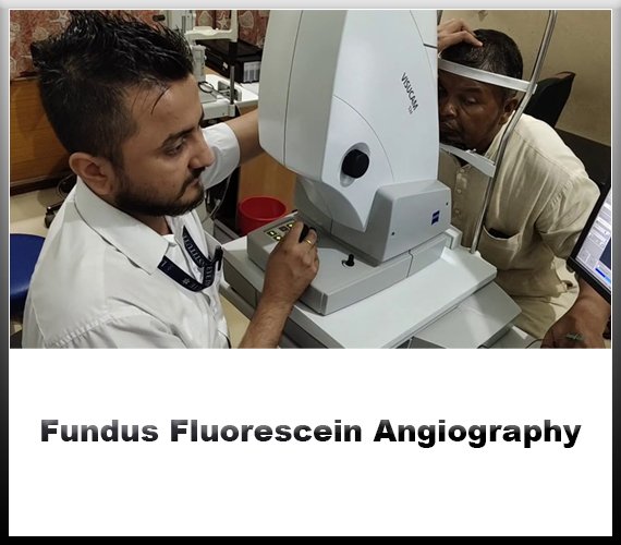

What is Fundus Fluorescein Angiography?
Fundus fluorescein angiography (FFA) is a diagnostic procedure used to evaluate the blood circulation in the back of the eye, particularly the retina and choroid. It employs a special dye called fluorescein, which is injected into a vein in the arm. As the dye circulates through the bloodstream, it highlights the blood vessels in the eye when illuminated by a specific wavelength of light. This allows ophthalmologists to capture detailed images of the retina and choroid, helping them diagnose and monitor various eye conditions.
The procedure typically begins with the patient's pupils being dilated using eye drops to ensure a clear view of the back of the eye. After the pupils are dilated, a small amount of fluorescein dye is injected into a vein, usually in the arm. As the dye travels through the bloodstream, it reaches the blood vessels of the eye within seconds. A special camera equipped with filters to
capture the fluorescent light emitted by the dye is then used to take sequential images of the retina and choroid.
FFA provides valuable information about the health and function of the blood vessels in the eye. It can help detect abnormalities such as leaking blood vessels, blockages, abnormal growth of blood vessels, and areas of poor blood flow. These findings are crucial in the diagnosis and management of various retinal and choroidal diseases, including diabetic retinopathy, macular degeneration, retinal vein occlusion, and uveitis.
One of the key advantages of FFA is its ability to capture dynamic changes in the circulation of the eye. By taking images at different time points after the dye injection, ophthalmologists can observe how the dye moves through the blood vessels and identify any abnormal patterns or areas of leakage. This information is essential for planning appropriate treatment strategies and monitoring the response to therapy over time.
Despite its benefits, FFA does have some limitations and risks. The dye injection may cause mild side effects such as nausea, vomiting, or allergic reactions in some patients. Additionally, the procedure requires the use of dilating eye drops, which can temporarily blur vision and increase sensitivity to light. In rare cases, more serious complications such as infection or damage to the eye may occur.
fundus fluorescein angiography is a valuable diagnostic tool in the field of ophthalmology, allowing for the detailed assessment of retinal and choroidal circulation. By providing real-time information about blood flow and vascular abnormalities, FFA plays a crucial role in the diagnosis and management of various eye diseases, ultimately helping to preserve and restore vision in affected individuals.


Frequently Asked Questions
Choose Jain Eye Hospital for expert eye care. With cutting-edge technology and compassionate specialists, we ensure optimal vision health for you and your loved ones.
Over time, cataracts will harm your vision. Cataract surgery can bring back your vision. However, a possible complication of cataract surgery is an "after-cataract." An "after-cataract" happens when part of the natural lens that is purposely not taken out during cataract surgery becomes cloudy and blurs your eyesight.
How painful is cataract surgery? Most people feel little or no pain during or after cataract surgery. You'll receive a topical anesthetic (eye drops) to numb your eye during the surgery. Shortly after surgery, your eye may feel gritty or slightly tender, but over-the-counter pain medicine should improve this.
Many factors can induce inflammation of ocular surface, including DED, pathogens, and allergic reactions. Acute inflammation is usually beneficial, as it promotes the process of healing. But chronic inflammation may result in cell damage or even cell death.
Diagnosis and treatment of inflammatory eye diseases are important. They can cause permanent damage to the eyes and vision loss that cannot be reversed. If you notice any of the signs or symptoms of inflammatory eye disease, make an appointment to see your eye doctor right away for a complete eye exam.
We are the best
Welcome to Jain Eye Hospital, where your vision is our priority. With a legacy of excellence spanning over three decades, we are committed to providing world-class eye care services with compassion, expertise, and cutting-edge technology.
- expert in eye care.
- State-of-the-art facilities.
- Comprehensive services.
- Patient-centered approach.
Our Services


Diabetic Retinopathy Management
Such as photocoagulation or panretinal photocoagulation, halts blood and fluid leakage and shrinks abnormal vessels in the eye.
Read More
Ocular Inflammation
Ocular inflammation is inflammation of the eye, which can be caused by many factors, including pathogens, allergic reactions, and DED.
Read More
Visual Field Analysis
A visual field test, also known as a perimetry test, is a painless eye exam that measures the area of vision and how sensitive it is in different parts.It can detect blind spots in your vision.
Read More
We are available 24/7
We are Always Ready For A Challenge.
Satisfied Patients
Patient satisfaction is an attitude. Though it does not ensure that the patient will remain loyal to the doctor or the hospital, it is still a strong.
Awards
Highly skilled medical professionals offering specialized care and advanced treatment modalities.
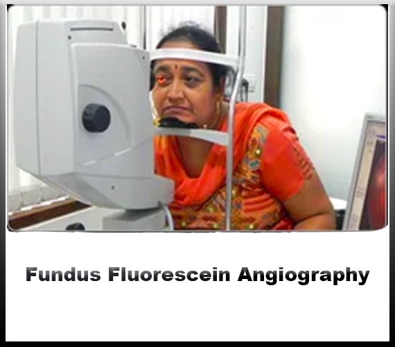
How do you Fundus Fluorescein Angiography?
Fundus Fluorescein Angiography (FFA) is a valuable diagnostic tool used in ophthalmology to assess the circulation of the retina and choroid. It involves the intravenous injection of a fluorescent dye, typically sodium fluorescein, followed by capturing sequential images of the retina using a specialized camera equipped with filters that can detect the fluorescence emitted by the dye. The resulting images provide detailed information about the blood flow and vascular integrity of the retina, aiding in the diagnosis and management of various ocular conditions.
The procedure begins with the preparation of the patient and the equipment. The patient's medical history, including any allergies or contraindications to fluorescein dye, is reviewed to ensure the safety of the procedure. After obtaining informed consent, the patient's pupils are dilated using topical ophthalmic drops to allow for optimal visualization of the retina.
Next, the fluorescein dye is injected into a vein, typically in the arm, using a sterile technique. The dye rapidly circulates through the bloodstream and reaches the retinal vessels within seconds. As the dye perfuses through the retinal vasculature, the specialized camera captures a series of images at specific time intervals, typically ranging from seconds to minutes, depending on the clinical indication and the information required.
During image acquisition, the camera emits a blue light, which excites the fluorescein dye, causing it to fluoresce and emit a greenish-yellow light. This fluorescence allows for the visualization of the retinal vessels and any abnormalities in their architecture or perfusion patterns. The sequential images provide dynamic information about the circulation, including the arterial and venous phases, as well as the presence of any leakage or pooling of dye, which can indicate areas of vascular compromise or pathology.
Interpretation of FFA images requires careful analysis by an experienced ophthalmologist or retina specialist. They assess the integrity of the retinal vasculature, identify any areas of non-perfusion, neovascularization, or macular edema, and correlate these findings with the patient's clinical presentation and other diagnostic tests. FFA is particularly useful in the diagnosis and management of retinal vascular diseases such as diabetic retinopathy, age-related macular degeneration, retinal vein occlusions, and inflammatory conditions affecting the retina.
Fundus Fluorescein Angiography is a valuable imaging technique in ophthalmology that provides dynamic information about the retinal circulation and aids in the diagnosis and management of various ocular conditions. Through the intravenous injection of fluorescein dye and sequential imaging of the retina, FFA allows for the visualization of vascular abnormalities and helps guide treatment decisions to optimize visual outcomes for patients.