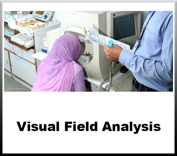

What is Visual Field Analysis?
Visual field analysis, also known as perimetry, is a diagnostic procedure used to assess the full horizontal and vertical range of vision of an individual. It is a crucial tool in ophthalmology and neurology for detecting and monitoring various conditions affecting the visual pathway, such as glaucoma, optic nerve disorders, retinal diseases, and neurological conditions like brain tumors or strokes.
The human visual field is the total area in which objects can be seen while the eye is focused on a central point. Visual field analysis measures this area by systematically testing the patient's ability to perceive visual stimuli presented at different locations within their field of view. This assessment helps in identifying any abnormalities or defects in the visual field, which may indicate underlying pathology.
The most common method of visual field analysis involves automated perimetry, where the patient sits in front of a concave dome or a
computerized device with a screen. During the test, tiny points of light are presented at various locations within the patient's visual field, and the patient is instructed to respond whenever they see a light flash. The responses are recorded, creating a map of the patient's visual field sensitivity.
Several parameters are measured during visual field analysis, including mean deviation, pattern standard deviation, and visual field index, which provide information about the overall sensitivity and uniformity of the visual field. These measurements help in quantifying any abnormalities and tracking changes over time.
Visual field analysis is particularly valuable in the diagnosis and management of glaucoma, a progressive optic neuropathy characterized by damage to the optic nerve and visual field loss. By regularly monitoring the visual field of glaucoma patients, clinicians can detect early signs of progression and adjust treatment strategies accordingly to preserve remaining vision.
In addition to glaucoma, visual field analysis is also used in the evaluation of other optic nerve and retinal disorders, such as optic neuritis, retinitis pigmentosa, and diabetic retinopathy. Furthermore, it plays a vital role in assessing visual function in patients with neurological conditions affecting the visual pathway, including brain tumors, multiple sclerosis, and stroke.
Overall, visual field analysis is an indispensable tool in the diagnosis, management, and monitoring of various ocular and neurological conditions affecting visual function. By providing quantitative data about the integrity of the visual field, it aids clinicians in making informed decisions regarding treatment and patient care.


Frequently Asked Questions
Choose Jain Eye Hospital for expert eye care. With cutting-edge technology and compassionate specialists, we ensure optimal vision health for you and your loved ones.
Over time, cataracts will harm your vision. Cataract surgery can bring back your vision. However, a possible complication of cataract surgery is an "after-cataract." An "after-cataract" happens when part of the natural lens that is purposely not taken out during cataract surgery becomes cloudy and blurs your eyesight.
How painful is cataract surgery? Most people feel little or no pain during or after cataract surgery. You'll receive a topical anesthetic (eye drops) to numb your eye during the surgery. Shortly after surgery, your eye may feel gritty or slightly tender, but over-the-counter pain medicine should improve this.
Many factors can induce inflammation of ocular surface, including DED, pathogens, and allergic reactions. Acute inflammation is usually beneficial, as it promotes the process of healing. But chronic inflammation may result in cell damage or even cell death.
Diagnosis and treatment of inflammatory eye diseases are important. They can cause permanent damage to the eyes and vision loss that cannot be reversed. If you notice any of the signs or symptoms of inflammatory eye disease, make an appointment to see your eye doctor right away for a complete eye exam.
We are the best
Welcome to Jain Eye Hospital, where your vision is our priority. With a legacy of excellence spanning over three decades, we are committed to providing world-class eye care services with compassion, expertise, and cutting-edge technology.
- expert in eye care.
- State-of-the-art facilities.
- Comprehensive services.
- Patient-centered approach.
Our Services


Diabetic Retinopathy Management
Such as photocoagulation or panretinal photocoagulation, halts blood and fluid leakage and shrinks abnormal vessels in the eye.
Read More
Ocular Inflammation
Ocular inflammation is inflammation of the eye, which can be caused by many factors, including pathogens, allergic reactions, and DED.
Read More
Visual Field Analysis
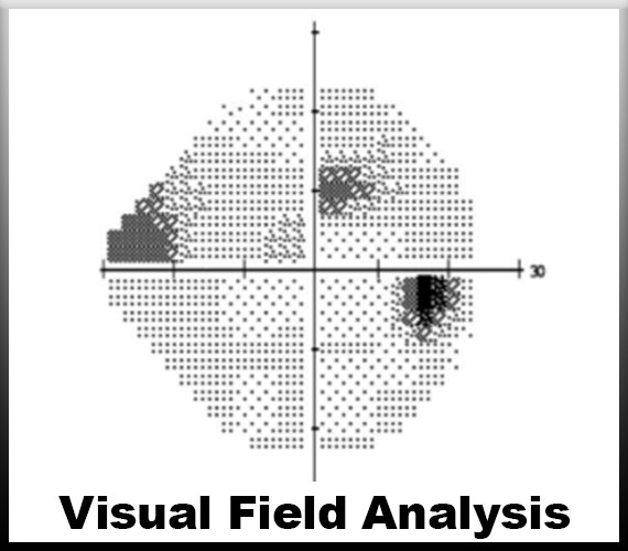
A visual field test, also known as a perimetry test, is a painless eye exam that measures the area of vision and how sensitive it is in different parts.It can detect blind spots in your vision.
Read More
We are available 24/7
We are Always Ready For A Challenge.
Satisfied Patients
Patient satisfaction is an attitude. Though it does not ensure that the patient will remain loyal to the doctor or the hospital, it is still a strong.
Awards
Highly skilled medical professionals offering specialized care and advanced treatment modalities.
How do you Analyse visual fields?
Analyzing visual fields involves assessing the extent and quality of a person's peripheral vision, crucial for detecting abnormalities such as glaucoma or neurological disorders. Various methods exist, with the Goldman perimeter and automated perimetry being common.
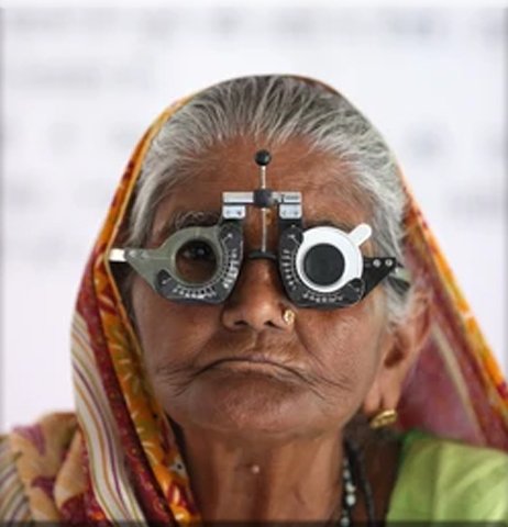
Goldman perimeter: This traditional method employs a bowl-shaped instrument where a patient fixes their gaze while lights of varying intensities are projected into their visual field. The patient signals when they detect the light, allowing the examiner to map out their peripheral vision. Results are plotted on a grid, indicating areas of reduced sensitivity.
Automated perimetry: Utilizing advanced technology, automated perimetry involves a computerized instrument that presents stimuli at precise locations within the visual field. The patient responds to stimuli, typically by
pressing a button, and the instrument records their responses, generating a detailed map of their visual field sensitivity.
Interpretation: Analysis of visual fields requires careful examination of the resulting maps. Common parameters include mean deviation (average deviation from normal sensitivity), pattern standard deviation (variance from an expected pattern of sensitivity), and visual field indices. Comparison with age-matched norms aids in identifying abnormalities.
Considerations: Factors such as patient cooperation, learning effects, and artifacts must be considered during analysis. Repeat testing may be necessary to confirm results and monitor progression.
Clinical significance: Abnormalities in visual fields may indicate various conditions, including glaucoma, optic nerve damage, retinal disorders, or neurological diseases. Early detection through visual field analysis enables timely intervention and management, preserving vision and quality of life.
visual field analysis is a fundamental aspect of ophthalmic and neurological assessment, providing valuable insights into a patient's peripheral vision and overall visual health. Both traditional and automated methods offer reliable means of mapping visual fields, aiding in the diagnosis and management of a wide range of ocular and neurological conditions.